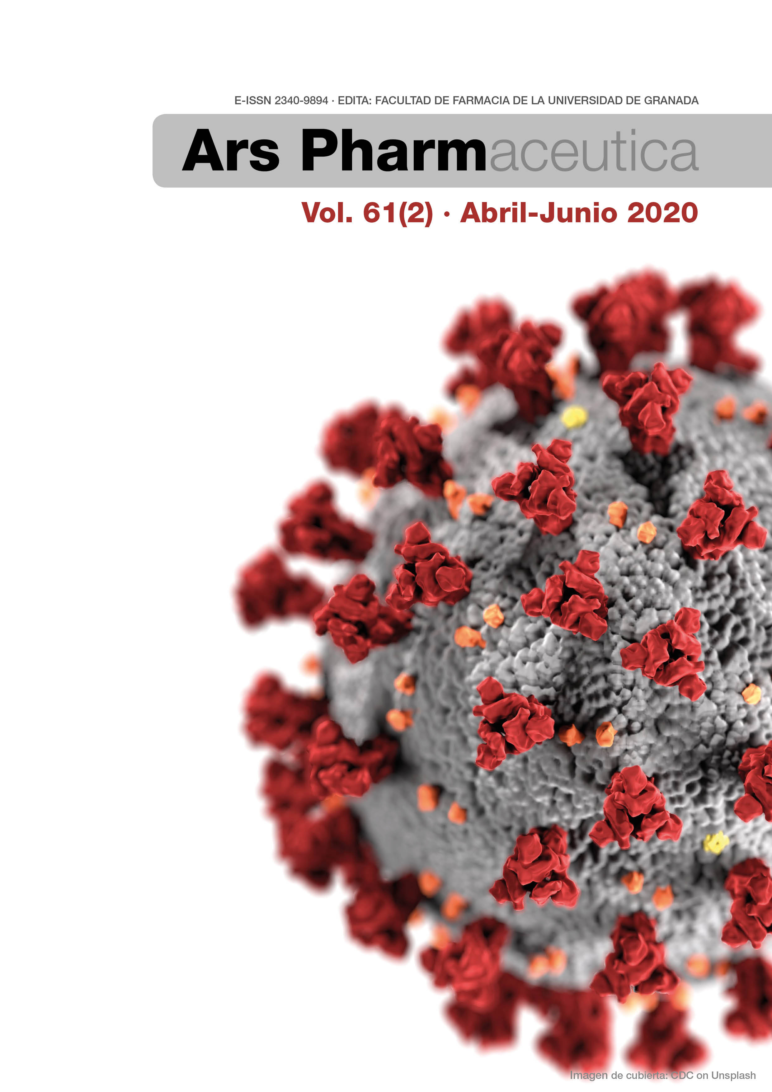
SARS-CoV-2 y pandemia de síndrome respiratorio agudo (COVID-19)
Palabras clave:
SARS-CoV-2, COVID-19, coronavirus.Resumen
Introducción: En diciembre de 2019, se detectaron los primeros casos de enfermedad respiratoria causada por un coronavirus emergente, al que se denominó SARS-CoV-2, que en los primeros meses de 2020 se ha extendido por todo el mundo con características de pandemia.
Método: Se examinaron las publicaciones más relevantes en relación con los objetivos de la revisión.
Resultados: La enfermedad, conocida como COVID-19, cursa con tos, fiebre y dificultad respiratoria. Las formas más graves, que afectan principalmente a personas de edad avanzada y con determinadas comorbilidades, se manifiestan por afectación de la función respiratoria, que requiere ventilación mecánica, y síndrome de respuesta inflamatoria sistémica, que puede conducir a un choque séptico con fallo multiorgánico, y altas tasas de mortalidad. En esta revisión se examina el estado actual de conocimientos sobre las características y origen del SARS-CoV-2, su replicación, y la patogénesis, clínica, diagnóstico, tratamiento y prevención de COVID-19.
Conclusiones: Las características del SARS-CoV-2 y la clínica de COVID-19 son bien conocidas. La PCR es la técnica de referencia para el diagnóstico de laboratorio; se dispone de ensayos para detección de antígenos y de anticuerpos, con margen de optimización. Los protocolos de tratamiento incluyen la corrección de la respuesta inflamatoria sistémica y administración de agentes antivirales. Existen vacunas en desarrollo.
Descargas
Citas
Tyrrell DAJ, Bynoe ML. Cultivation of a novel type of common-cold virus in organ cultures. Brit Med J. 1965;1(5448):1467-70. doi.org/10.1136/bmj.1.5448.1467.
Hamre D, Procknow JJ. A new virus isolated from the human respiratory tract. Proc Soc Exp Biol Med. 1966;121(1):190-3. doi.org/10.3181/00379727-121-30734.
Kahn JS, McIntosh K. History and recent advances in coronavirus discovery. Pediatr Infect Dis J. 2005;24(11 Suppl):S223-7. doi.org/10.1097/01.inf.0000188166.17324.60.
Anónimo. Coronaviruses. Nature. 1968;220:650.
International Commitee of taxonomy of Viruses. Taxonomy; 2020. https://talk.ictvonline.org/taxonomy/ (acceso 04/04/2020).
Wildy P. Classification and nomenclature of viruses. First Report of the International Committee on Nomenclature of Viruses. Monographs in virology no. 5. Basel: Karger; 1971.
McIntosh K. Coronavirus. In: Richman DD, Whitley RJ, Hayden FG, editors. Clinical virology. Washington: ASM Press; 2002. p. 1087-96.
Gadsby NJ, Templeton KE. Coronaviruses. In: Jorgensen JH, Pfaller MA, Carroll KC, Funke G, Landry ML, Richter SS, Warnock DW, editors. Manual of clinical microbiology. Washington: ASM Press; 2015. p. 1565-83.
Fehr AR, Channappanavar R, Perlman S. Middle East Respiratory Syndrome: Emergence of a pathogenic human coronavirus. Annu Rev Med. 2017;68:387-99. doi.org/10.1146/annurev-med-051215-031152.
Graham RL, Donaldson EF, Baric RS. A decade after SARS: strategies for controlling emerging coronaviruses. Nat Rev Microbiol. 2013;11(12):836-48. doi.org/10.1038/nrmicro3143.
Lim YX, Ng YL, Tam JP, Liu DX. Human Coronaviruses: A Review of Virus-Host Interactions. Diseases. 2016;4(3). pii:E26. doi.org/10.3390/diseases4030026.
Zhou Y, Jiang S, Du L. Prospects for a MERS-CoV spike vaccine. Expert Rev Vaccines. 2018;17(8):677-86. doi.org/10.1080/14760584.2018.1506702.
Mackay M, Arden KE. MERS coronavirus: diagnostics, epidemiology and transmisión. Virol J. 2015:12:222. doi.org/10.1186/s12985-015-0439-5.
Comisión Municipal de Salud de Wuhan. Sobre la situación actual de neumonía en nuestra ciudad (en chino). Wuhan; 2019. http://wjw.wuhan.gov.cn/front/web/showDetail/2019123108989 (acceso 04/04/2020)
ECDC (European Centre for Disease Prevention and Control). Cluster of pneumonia cases caused by a novel coronavirus, Wuhan, China. 26 January 2020.
https://www.ecdc.europa.eu/sites/default/files/documents/Risk-assessment-pneumonia-Wuhan-China-26-Jan-2020_0.pdf (acceso 04/04/2020)
Wu F, Zhao S, Yu B, Chen YM, Wang W, Song ZG, Hu Y, Tao ZW, Tian JH, Pei YY, Yuan ML, Zhang YL, Dai FH, Liu Y, Wang QM, Zheng JJ, Xu L, Holmes EC, Zhang YZ. A new coronavirus associated with human respiratory disease in China. Nature. 2020;579(7798):265-9. doi.org/10.1038/s41586-020-2008-3.
Zhu N, Zhang D, Wang W, Li X, Yang B, Song J, Zhao X, Huang B, Shi W, Lu R, Niu P, Zhan F, Ma X, Wang D, Xu W, Wu G, Gao GF, Tan W. A novel coronavirus from patients with pneumonia in China, 2019. N Engl J Med. 2020;382(8):727-733. doi.org/10.1056/NEJMoa2001017.
Coronaviridae Study Group of the International Committee on Taxonomy of Viruses. The species Severe acute respiratory syndrome-related coronavirus: classifying 2019-nCoV and naming it SARS-CoV-2. Nat Microbiol. 2020;5(4):536-44. doi.org/10.1038/s41564-020-0695-z.
WHO. WHO Director-General’s remarks at the media briefing on 2019-nCoV. 2020. https://www.who.int/dg/speeches/detail/who-director-general-s-remarks-at-the-media-briefing-on-2019-ncov-on-11-february-2020 (acceso 04/04/2020).
Alocución de apertura del Director General de la OMS en la rueda de prensa sobre la COVID-19 celebrada el 11 de marzo de 2020. https://www.who.int/es/dg/speeches/detail/who-director-general-s-opening-remarks-at-the-media-briefing-on-covid-19---11-march-2020 (acceso 04/04/2020).
WHO. Coronavirus disease 2019 (COVID-19) Situation Report – 74. 2020. https://www.who.int/docs/default-source/coronaviruse/situation-reports/20200403-sitrep-74-covid-19-mp.pdf?sfvrsn=4e043d03_4 (acceso 04/04/2020).
ViralZone. Betacoronavirus. 2020. https://viralzone.expasy.org/764 (acceso 05/04/2020).
Masters PS. The molecular biology of coronaviruses. Adv Virus Res. 2006;66:193-292. doi.org/10.1016/S0065-3527(06)66005-3.
Ou X, Liu Y, Lei X, Li P, Mi D, Ren L, Guo L, Guo R, Chen T, Hu J, Xiang Z, Mu Z, Chen X, Chen J, Hu K, Jin Q, Wang J, Qian Z. Characterization of spike glycoprotein of SARS-CoV-2 on virus entry and its immune cross-reactivity with SARS-CoV. Nat Commun. 2020;11(1):1620. doi.org/10.1038/s41467-020-15562-9.
Neuman BW, Kiss G, Kunding AH, Bhella D, Baksh MF, Connelly S, Droese B, Klaus JP, Makino S, Sawicki SG, Siddell SG, Stamou DG, Wilson IA, Kuhn P, Buchmeier MJ. A structural analysis of M protein in coronavirus assembly and morphology. J Struct Biol. 2011;174(1):11-22. doi.org/10.1016/j.jsb.2010.11.021.
Nal B, Chan C, Kien F, Siu L, Tse J, Chu K, Kam J, Staropoli I, Crescenzo-Chaigne B, Escriou N, van der Werf S, Yuen KY, Altmeyer R. Differential maturation and subcellular localization of severe acute respiratory síndrome coronavirus surface proteins S, M and E. J Gen Virol. 2005;86(Pt 5):1423-34. doi.org/10.1099/vir.0.80671-0.
Chen Y, Liu Q, Guo D. Emerging coronaviruses: Genome structure, replication, and pathogenesis. J Med Virol. 2020;92(4):418-23. doi.org/10.1002/jmv.25681.
Tang X, Wu C, Xiang Li, Yuhe Song, Xinmin Yao, Xinkai Wu, Yuange Duan, Hong Zhang, Yirong Wang, Zhaohui Qian, Jie Cui, Jian Lu, On the origin and continuing evolution of SARS-CoV-2. Nat Sci Rev. 2020. nwaa036. doi.org/10.1093/nsr/nwaa036.
Lan J, Ge J, Yu J, Shan S, Zhou H, Fan S, Zhang Q, Shi X, Wang Q, Zhang L, Wang X. Structure of the SARS-CoV-2 spike receptor-binding domain bound to the ACE2 receptor. Nature. 2020. doi.org/10.1038/s41586-020-2180-5.
Cao YC, Deng QX, Dai SX. Remdesivir for severe acute respiratory síndrome coronavirus 2 causing COVID-19: An evaluation of the evidence. Travel Med Infect Dis. 2020:101647. doi.org/10.1016/j.tmaid.2020.101647.
Hoffmann M, Kleine-Weber H, Schroeder S, Krüger N, Herrler T, Erichsen S, Schiergens TS, Herrler G, Wu NH, Nitsche A, Müller MA, Drosten C, Pöhlmann S. SARS-CoV-2 Cell entry depends on ACE2 and TMPRSS2 and is blocked by a clinically proven protease inhibitor. Cell. 2020 Mar 4. pii: S0092-8674(20)30229-4. doi.org/10.1016/j.cell.2020.02.052.
Chen YW, Yiu CB, Wong KY. Prediction of the SARS-CoV-2 (2019-nCoV) 3C-like protease (3CL (pro)) structure: virtual screening reveals velpatasvir, ledipasvir, and other drug repurposing candidates. F1000Res. 2020;9:129. doi.org/10.12688/f1000research.22457.1.
Park WB, Kwon NJ, Choi SJ, Kang CK, Choe PG, Kim JY, Yun J, Lee GW, Seong MW, Kim NJ, Seo JS, Oh MD. Virus isolation from the first patient with SARS-CoV-2 in Korea. J Korean Med Sci. 2020;35(7):e84. doi.org/10.3346/jkms.2020.35.e84.
Banerjee A, Kulcsar K, Misra V, Frieman M, Mossman K. Bats and coronaviruses. Viruses. 2019;11(1). pii: E41. doi.org/10.3390/v11010041.
Zhou P, Yang XL, Wang XG, Hu B, Zhang L, Zhang W, Si HR, Zhu Y, Li B, Huang CL, Chen HD, Chen J, Luo Y, Guo H, Jiang RD, Liu MQ, Chen Y, Shen XR, Wang X, Zheng XS, Zhao K, Chen QJ, Deng F, Liu LL, Yan B, Zhan FX, Wang YY, Xiao GF, Shi ZL. A pneumonia outbreak associated with a new coronavirus of probable bat origin. Nature. 2020;579(7798):270-3. doi.org/10.1038/s41586-020-2012-7.
Guo YR, Cao QD, Hong ZS, Tan YY, Chen SD, Jin HJ, Tan KS, Wang DY, Yan Y. The origin, transmission and clinical therapies on coronavirus disease 2019 (COVID-19) outbreak - an update on the status. Mil Med Res. 2020;7(1):11. doi.org/10.1186/s40779-020-00240-0.
Zhang C, Zheng W, Huang X, Bell EW, Zhou X, Zhang Y. Protein structure and sequence reanalysis of 2019-nCoV genome refute snakes as its intermediate host and the unique similarity between its spike protein insertions and HIV-1. J Proteome Res. 2020;19(4):1351-60. doi.org/10.1021/acs.jproteome.0c00129.
Menachery VD, Yount BL Jr, Debbink K, Agnihothram S, Gralinski LE, Plante JA, Graham RL, Scobey T, Ge XY, Donaldson EF, Randell SH, Lanzavecchia A, Marasco WA, Shi ZL, Baric RS. A SARS-like cluster of circulating bat coronaviruses shows potential for human emergence. Nat Med. 2015;21(12):1508-13. doi.org/10.1038/nm.3985.
Andersen KG, Rambaut A, Lipkin WI, Holmes CE, Garry RF. The proximal origin of SARS-CoV-2. Nat Med 2020. doi.org/10.1038/s41591-020-0820-9.
Huang C, Liu WJ, Xu W, Jin T, Zhao Y, Song J, Shi Y, Ji W, Jia H, Zhou Y, Wen H, Zhao H, Liu H, Li H, Wang Q, Wu Y, Wang L, Liu D, Liu G, Yu H, Holmes EC, Lu L, Gao GF. A bat-derived putative cross-family recombinant coronavirus with a reovirus gene. PLoS Pathog. 2016;12(9):e1005883. doi.org/10.1371/journal.ppat.1005883.
Obameso JO, Li H, Jia H, Han M, Zhu S, Huang C, Zhao Y, Zhao M, Bai Y, Yuan F, Zhao H, Peng X, Xu W, Tan W, Zhao Y, Yuen KY, Liu WJ, Lu L, Gao GF. The persistent prevalence and evolution of cross-family recombinant coronavirus GCCDC1 among a bat population: a two-year follow-up. Sci China Life Sci. 2017;60(12):1357-1363. doi.org/10.1007/s11427-017-9263-6.
Chafekar A, Fielding BC. MERS-CoV: Understanding the latest human coronavirus threat. Viruses. 2018;10(2). pii: E93. doi.org/10.3390/v10020093.
Prompetchara E, Ketloy C, Palaga T. Immune responses in COVID-19 and potential vaccines: Lessons learned from SARS and MERS epidemic. Asian Pac J Allergy Immunol. 2020;38(1):1-9. doi.org/10.12932/AP-200220-0772.
Kutter JS, Spronken MI, Fraaij PL, Fouchier RA, Herfst S. Transmission routes of respiratory viruses among humans. Curr Opin Virol. 2018;28:142-51. doi.org/10.1016/j.coviro.2018.01.001.
Ministerio de Sanidad. Centro de Coordinación de Alertas y Emergencias Sanitarias. Enfermedad por coronavirus, COVID-19. Actualización 4 abril 2020. https://www.mscbs.gob.es/profesionales/saludPublica/ccayes/alertasActual/nCov-China/documentos/20200404_ITCoronavirus.pdf (acceso 13/04/2020).
van Doremalen N, Bushmaker T, Morris DH, Holbrook MG, Gamble A, Williamson BN, Tamin A, Harcourt JL, Thornburg NJ, Gerber SI, Lloyd-Smith JO, de Wit E, Munster VJ. Aerosol and surface stability of SARS-CoV-2 as compared with SARS-CoV-1. N Engl J Med. 2020. doi.org/10.1056/NEJMc2004973.
Park M, Cook AR, Lim JT, Sun Y, Dickens BL. A systematic review of COVID-19 epidemiology based on current evidence. J Clin Med. 2020;9(4). pii: E967. doi.org/10.3390/jcm9040967.
Zou L, Ruan F, Huang M, Liang L, Huang H, Hong Z, Yu J, Kang M, Song Y, Xia J, Guo Q, Song T, He J, Yen HL, Peiris M, Wu J. SARS-CoV-2 Viral Load in Upper Respiratory Specimens of Infected Patients. N Engl J Med. 2020;382(12):1177-9. doi.org/10.1056/NEJMc2001737.
Wölfel R, Corman VM, Guggemos W, Seilmaier M, Zange S, Müller MA, Niemeyer D, Jones TC, Vollmar P, Rothe C, Hoelscher M, Bleicker T, Brünink S, Schneider J, Ehmann R, Zwirglmaier K, Drosten C, Wendtner C. Virological assessment of hospitalized patients with COVID-2019. Nature. 2020. doi.org/10.1038/s41586-020-2196-x.
Lin L, Lu L, Cao W, Li T. Hypothesis for potential pathogenesis of SARS-CoV-2 infection-a review of immune changes in patients with viral pneumonia. Emerg Microbes Infect. 2020;9(1):727-32. doi.org/10.1080/22221751.2020.1746199.
Wong SH, Lui RN, Sung JJ. Covid-19 and the Digestive System. J Gastroenterol Hepatol. 2020. doi.org/10.1111/jgh.15047.
Siddiqi HK, Mehra MR. COVID-19 Illness in native and immunosuppressed states: a clinical-therapeutic staging proposal. J Heart Lung Transplant. 2020. doi.org/10.1016/j.healun.2020.03.012.
Li X, Geng M, Peng Y, Men L, Lu S. Molecular immune pathogenesis and diagnosis of COVID-19. J Pharm Anal. 2020. doi.org/10.1016/j.jpha.2020.03.001
Jaume M, Yip MS, Cheung CY, Leung HL, Li PH, Kien F, Dutry I, Callendret B, Escriou N, Altmeyer R, Nal B, Daëron M, Bruzzone R, Peiris JS. Anti-severe acute respiratory syndrome coronavirus spike antibodies trigger infection of human immune cells via a pH- and cysteine protease-independent FcγR pathway. J Virol. 2011;85(20):10582-97. doi.org/10.1128/JVI.00671-11.
Shi Y, Wang Y, Shao C, Huang J, Gan J, Huang X, Bucci E, Piacentini M, Ippolito G, Melino G. COVID-19 infection: the perspectives on immune responses. Cell Death Differ. 2020. doi.org/10.1038/s41418-020-0530-3.
Drew W, Wilson DV, Sapey E. Inflammation and neutrophil immunosenescence in health and disease: Targeted treatments to improve clinical outcomes in the elderly. Exp Gerontol. 2018;105:70-7. doi.org/10.1016/j.exger.2017.12.020.
Thomas R, Wang W, Su DM. Contributions of age-related thymic involution to immunosenescence and inflammaging. Immun Ageing. 2020;17:2. doi.org/10.1186/s12979-020-0173-8.
Oh SJ, Lee JK, Shin OS. Aging and the immune system: the impact of immunosenescence on viral infection, immunity and vaccine immunogenicity. Immune Netw. 2019;19(6):e37. doi.org/10.4110/in.2019.19.e37.
Monneret G, Gossez M, Venet F. Sepsis and immunosenescence: closely associated in a vicious circle. Aging Clin Exp Res. 2019. doi.org/10.1007/s40520-019-01350-z.
Ciaglia E, Vecchione C, Puca AA. COVID-19 infection and the predictive ACE2 soluble levels: the favourable protection of children and women. Front Pediatr. 2020. doi.org/10.3389/fped.2020.00206
Passanisi S, Lombardo F, Salzano G, Pajno GB. Are children most of the submerged part of SARS-CoV-2 iceberg? Front Pediatr. 2020. doi.org/10.3389/fped.2020.00213.
Sociedad Española de Enfermedades Infecciosas y Microbiología Clínica (SEIMC). Recomendaciones institucionales. Documento de posicionamiento de la SEIMC sobre el diagnóstico microbiólogo de COVID-19. 2020.
https://seimc.org/contenidos/documentoscientificos/recomendaciones/seimc-rc-2020-Posicionamiento_SEIMC_diagnostico_microbiologico_COVID19.pdf (acceso 13/04/2020).
Bell JJ, Selvarangan R. Evaluation of the Alere I influenza A&B nucleic acid amplification test by use of respiratory specimens collected in viral transport medium. J Clin Microbiol. 2014;52(11):3992-5. doi.org/10.1128/JCM.01639-14.
Patel R, Babady E, Theel ES, Storch GA, Pinsky BA, St George K, Smith TC, Bertuzzi S. Report from the American Society for Microbiology COVID-19 International Summit, 23 March 2020: value of diagnostic testing for SARS-CoV-2/COVID-19. mBio. 2020;11(2). pii: e00722-20. doi.org/10.1128/mBio.00722-20.
Ministerio de Sanidad. Centro de Coordinación de Alertas y Emergencias Sanitarias. Procedimiento de actuación frente a casos de infección por el nuevo coronavirus (SARS-CoV-2). Actualizado a 11 de abril de 2020. https://www.mscbs.gob.es/en/profesionales/saludPublica/ccayes/alertasActual/nCov-China/documentos/Procedimiento_COVID_19.pdf (acceso 14/04/2020).
Ahn DG, Shin HJ, Kim MH, Lee S, Kim HS, Myoung J, Kim BT, Kim SJ. Current status of epidemiology, diagnosis, therapeutics, and vaccines for novel coronavirus disease 2019 (COVID-19). J Microbiol Biotechnol. 2020;30(3):313-24. doi.org/10.4014/jmb.2003.03011.
Agencia Española de Medicamentos y Productos Sanitarios (AEMPS). Tratamientos disponibles para el manejo de la infección respiratoria por SARS-CoV-2. Actualizado a 28 de marzo de 2020. https://www.aemps.gob.es/laAEMPS/docs/medicamentos-disponibles-SARS-CoV-2-28-3-2020.pdf?x38929 (acceso 14/04/2020).
Vincent MJ, Bergeron E, Benjannet S, Erickson BR, Rollin PE, Ksiazek TG, Seidah NG, Nichol ST. Chloroquine is a potent inhibitor of SARS coronavirus infection and spread. Virol J. 2005;2:69. doi.org/10.1186/1743-422X-2-69.
Hagen A. Antimicrobials and COVID-19: strategies for treating a pandemic. March 23, 2020. https://www.asm.org/Articles/2020/March/Antimicrobials-and-COVID-19-Treatment-Strategies-f (acceso 14/04/2020)
Gautret P, Lagier JC, Parola P, Hoang VT, Meddeb L, Mailhe M, Doudier B, Courjon J, Giordanengo V, Vieira VE, Dupont HT, Honoré S, Colson P, Chabrière E, La Scola B, Rolain JM, Brouqui P, Raoult D. Hydroxychloroquine and azithromycin as a treatment of COVID-19: results of an open-label non-randomized clinical trial. Int J Antimicrob Agents. 2020 Mar 20:105949. doi.org/10.1016/j.ijantimicag.2020.105949.
Beigelman A, Gunsten S, Mikols CL, Vidavsky I, Cannon CL, Brody SL, Walter MJ. Azithromycin attenuates airway inflammation in a noninfectious mouse model of allergic asthma. Chest. 2009;136(2):498-506. doi.org/10.1378/chest.08-3056.
Vos R, Vanaudenaerde BM, Verleden SE, Ruttens D, Vaneylen A, Van Raemdonck DE, Dupont LJ, Verleden GM. Anti-inflammatory and immunomodulatory properties of azithromycin involved in treatment and prevention of chronic lung allograft rejection. Transplantation. 2012;94(2):101-9. doi.org/10.1097/TP.0b013e31824db9da.
Caly L, Druce JD, Catton MG, Jans DA, Wagstaff KM. The FDA-approved Drug Ivermectin inhibits the replication of SARS-CoV-2 in vitro. Antiviral Res. 2020:104787. doi.org/10.1016/j.antiviral.2020.104787.
Khan S, Siddique R, Shereen MA, Ali A, Liu J, Bai Q, Bashir N, Xue M. The emergence of a novel coronavirus (SARS-CoV-2), their biology and therapeutic options. J Clin Microbiol. 2020. pii: JCM.00187-20. doi.org/10.1128/JCM.00187-20.
Enjuanes L, Zuñiga S, Castaño-Rodriguez C, Gutierrez-Alvarez J, Canton J, Sola I. Molecular basis of coronavirus virulence and vaccine development. Adv Virus Res. 2016;96:245-86. doi.org/10.1016/bs.aivir.2016.08.003.
Amanat F, Krammer F. SARS-CoV-2 Vaccines: Status Report. Immunity. 2020. pii: S1074-7613(20)30120-5.
doi.org/10.1016/j.immuni.2020.03.007.
Peeples L. News Feature: Avoiding pitfalls in the pursuit of a COVID-19 vaccine. Proc Natl Acad Sci U S A. 2020. pii: 202005456. doi.org/10.1073/pnas.2005456117.
Descargas
Publicado
Cómo citar
Número
Sección
Licencia
Derechos de autor 2020 Alfonso Ruiz-Bravo, María Jimenez-Varela

Esta obra está bajo una licencia internacional Creative Commons Atribución-NoComercial-CompartirIgual 4.0.
Los artículos que se publican en esta revista están sujetos a los siguientes términos en relación a los derechos patrimoniales o de explotación:
- Los autores/as conservarán sus derechos de autor y garantizarán a la revista el derecho de primera publicación de su obra, la cual se distribuirá con una licencia Creative Commons BY-NC-SA 4.0 que permite a terceros reutilizar la obra siempre que se indique su autor, se cite la fuente original y no se haga un uso comercial de la misma.
- Los autores/as podrán adoptar otros acuerdos de licencia no exclusiva de distribución de la versión de la obra publicada (p. ej.: depositarla en un archivo telemático institucional o publicarla en un volumen monográfico) siempre que se indique la fuente original de su publicación.
- Se permite y recomienda a los autores/as difundir su obra a través de Internet (p. ej.: en repositorios institucionales o en su página web) antes y durante el proceso de envío, lo cual puede producir intercambios interesantes y aumentar las citas de la obra publicada. (Véase El efecto del acceso abierto).