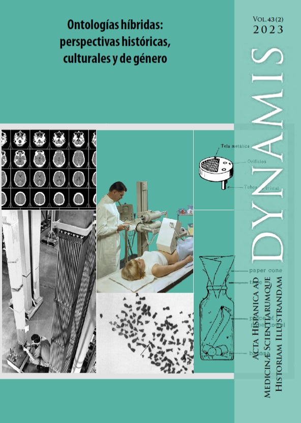
Digital brain imaging in the 1970s: building a hybrid technology
DOI:
https://doi.org/10.30827/dynamis.v43i2.29442Keywords:
visual cultures, computed tomography, digital picture, neurosciences, artistic research.Abstract
Computed tomography (CT), a technology based on the combination of radiology and computation, was used to scan a patient’s brain tumor in a London hospital in 1972. The patient’s head was introduced into a scanner, manufactured by the British record company EMI, to measure the amount of radiation absorbed by different points in the brain. The resulting image consisted of a digital tonal matrix that was materialized on newsprint, a cathode ray tube, and a Polaroid photograph. This article describes the production process of the CT image, a visual representation that would become, from the 1970s onwards, a common technology for contemporary neurosciences and a paradigm of visual representation in the transition from analog to digital. To this end, we analyze the implications of the radiographic image, invented at a time of the rise of theimage-movement in which chronophotographs, image sequences, and cinematography are framed. In this analysis, we focus on the scopic regime in which each image is found. The knowledge that led to construction of the device circulated between the clinic, industry, and laboratory. A variety of agents were involved in the construction of CT scans, including the computer, X-ray radiation, a team of electronic engineers, a group of neuroradiologists from Atkinson Marley Hospital, and the X-ray scanner, among others. We also report on the impact of several factors associated with the epistemology of CT imaging, such as the reinforcement of clinical diagnoses, the linking of the morphological to the psychic in relation to the brain, and the transition from image-motion to image-time.
Downloads
References
Ambrose, James. “Computerized transverse axial scanning (tomography): Part 2. Clinical application.” The British journal of radiology 46, n.º 552 (1973): 1023-1047. https://doi.org/10.1259/0007-1285-46-552-1023 DOI: https://doi.org/10.1259/0007-1285-46-552-1023
Anderson, Nancy A. y Dietrich, Michael. R. The educated eye: visual culture and pedagogy in the life sciences. New Hampshire: UPNE, 2012. DOI: https://doi.org/10.1349/ddlp.3554
Azoulay, Ariella. The Civil Contract of Photography. New York: Zone Books, 2008.
Beckmann, Elizabeth. C. “CT scanning the early days.” The British journal of radiology 79, n.º 937 (2006): 5-8. https://doi.org/10.1259/bjr/29444122 DOI: https://doi.org/10.1259/bjr/29444122
Benjamin, Walter. La obra de arte en la época de su reproductibilidad técnica. México: Ítaca, [1935] 2003.
Bhidé, Amar; Datar, Srikant y Stebbins, Katherine. “Case Histories of Significant Medical Advances: Development of Computed Tomography.” Harvard Business School Accounting & Management Unit Working Paper 20, n.º 4 (2021) (Revisado en julio de 2020). http://dx.doi.org/10.2139/ssrn.3429976 DOI: https://doi.org/10.2139/ssrn.3429976
Borden, William C. The Use of the Röntgen Ray by the Medical Department of the United States in the War with Spain. Washington: Government Printing Office, 1898.
Bosch, Enrique. “Sir Godfrey Newbold Hounsfield y la tomograf ía computada, su contribución a la medicina moderna.” Revista chilena de radiología 10, n.º 4 (2004): 183-185. http://dx.doi.org/10.4067/S0717-93082004000400007 DOI: https://doi.org/10.4067/S0717-93082004000400007
Bovery, Margaret y Glasser, Otto. Wilhelm Conrad Röntgen and the early history of the Roentgen rays. Estados Unidos: Norman Publishing, [1934] 1993.
Brea, José Luís. Las tres eras de la imagen: imagen-materia, film, e-image. Madrid: Akal, 2010.
Cartwright, Lisa. Screening the body: Tracing medicine’s visual culture. Minnesota: University of Minnesota Press, 1995.
Crary, Jonathan. Techniques of the observer: On vision and modernity in the nineteenth century. Cambridge: MIT press, 1990.
Curtis, Scott. “Photography and Medical Observation del capítulo”. The Educated Eye. Visual Culture and Pedagogy In the Life Sciences, ed. Nancy Anderson y Michael R. Dietrich (New Hampshire: Darmouth College Press, 2012), 67-86.
Daston, Lorraine y Galison, Peter. Objectivity. Nueva York: Zone Books, 2007. Delehanty, Megan. “Why images?” Medicine Studies 2, n.º 3 (2010): 161-173. https://doi.org/10.1007/s12376-010-0052-2 DOI: https://doi.org/10.1007/s12376-010-0052-2
Didi-Huberman, Georges. La imagen superviviente: historia del arte y tiempo de los fantasmas según Aby Warburg. Madrid: Abada, 2009.
Didi-Huberman, Georges. Ante la imagen: pregunta formulada a los fines de una historia del arte. Murcia: CENDEAC, 2010.
Dumit, Joseph. Picturing Personhood: Brain Scans and Biomedical Identity. Nueva Jersey: Princeton University Press, 2004. DOI: https://doi.org/10.1515/9780691236629
Ferragud, Carmel; Vidal, Antonio; Bertomeu, José. R. y Lucas, Rut. Documentación y metodología en Ciencias de la Salud. Valencia: Nau Llibres, 2017.
Fiorentini, Erna. “Induction of visibility: Reflections on histological slides, drawing visual hypotheses and aesthetic-epistemic actions”. History and philosophy of the life sciences, 35, n.º 3, 379-394 (2013). https://pubmed.ncbi.nlm.nih.gov/24779108/
Holtzmann Kevles, Bettyann. Naked to the bone. Medical Imaging in the Twentieth Century. Nueva Jersey: University Press, 1997. DOI: https://doi.org/10.1063/1.881857
Hounsfield, Godfrey Newbold. “Computerized transverse axial scanning (tomography): Part 1. Description of system.” The British journal of radiology 46, n.º 552 (1973): 1016-1022. https://doi.org/10.1259/0007-1285-46-552-1016 DOI: https://doi.org/10.1259/0007-1285-46-552-1016
Keller, Thomas M. “A Roentgen Centennial Legacy: The First Use of the X-Ray by the U.S. Military in the Spanish-American War.” Military medicine 162, n.º 8 (1997): 551-554. https://doi.org/10.1093/milmed/162.8.551 DOI: https://doi.org/10.1093/milmed/162.8.551
Krzywinski, Martin. “Scientific Data Visualization: Aesthetic of Diagrammatic Clarity.” En The Aesthetics of Scientific Data Representation: More than Pretty Pictures editado por Lotte Philipsen y Rikke S. Kjaergaard, 22-35. Nueva York: Routledge, 2018. DOI: https://doi.org/10.4324/9781315563411-3
Lamata Manuel, Ana. “Superrealistas: De la Contribución de los Rayos-X a la Visión y Presentación de la Realidad en el Arte a Comienzos del Siglo XX.” Tesis doctoral, Universidad Complutense de Madrid, 2010.
Lenander, Nanna. “X-ray Aesthetics. Radiographic Vision in The Magic Mountain and Painting, Photography, Film.” Tesis de Maestría, Universidad de Oslo, 2021.
J. Lanska, Douglas. “The dercum-muybridge collaboration and the study of pathologic gaits using sequential photography.” Journal of the History of the Neurosciences 25(1) (2016): 23-38. DOI: https://doi.org/10.1080/0964704X.2015.1070032
Latour, Bruno. La esperanza de pandora. Ensayos sobre la realidad de los estudios de la ciencia. Barcelona: Gedisa, [1999] 2001.
Mitchell, William J. T. La ciencia de la imagen: Iconología, cultura visual y estética de los medios. Madrid: Akal, [2015] 2019.
Moore, Kate. Las chicas del Radio. Lucharon por la justicia, pagaron con sus vidas. Madrid: Capitan Swing, 2018.
Muybridge, Eadweard. Horses and other animals in motion. Nueva York: Dover Publications, [1887] 1985.
Philipsen, Lotte y Kjaergaard, Rikke S. (eds.) The Aesthetics of Scientific Data Representation: More than Pretty Pictures. Nueva York: Routledge, 2018. DOI: https://doi.org/10.4324/9781315563411
Rego Robles, Miguel Ángel. The early drawings and prints of Santiago Ramón y Cajal: a visual epistemology of the neurosciences. European Journal of Anatomy, 23, n.º S1, 57-66 (2019).
Röntgen, Wilhelm C. “On a new kind of rays.” Science 3, n.º 59 (1896): 227-231.
https://doi.org/10.1126/science.3.59.227 DOI: https://doi.org/10.1126/science.3.59.227
Sansare, Kaustubh; Khanna, Vinit y Karjodkar, Freny. “Early victims of X-rays: atribute and current perception.” Dentomaxillofacial Radiology 40, n.º 2. (2011): 123-125. DOI: https://doi.org/10.1259/dmfr/73488299
https://dx.doi.org/10.1259%2Fdmfr%2F73488299
Secord, Anne. “Talbot´s first lens: Botanical vision as an exact science.” En William Henry Fox Talbot. Beyond photography, editado por M. Brusius. et al., 41-66. Yale: Yale University Press, 2013.
Spirt, Beverly A. y Randall, Patricia A. “Radiologic history exhibit. The Role of Women in Wartime Radiology.” RadioGraphics 15, n.º 3 (1995): 641-652. https://doi.org/10.1148/radiographics.15.3.7624569 DOI: https://doi.org/10.1148/radiographics.15.3.7624569
Susskind, Charles. “The invention of computed tomography.” En History of Technology editado por Rupert Hall y Norman Smith, 9-80. Londres: Mansell, 1981.
Van Dijck, José. The transparent body: A cultural analysis of medical imaging. Washington: University of Washington Press, 2011
Veldhuis, Djuke (2018). “Scientific Storytelling: Visualizing for Public Audiences.” En The Aesthetics of Scientific Data Representation: More than Pretty Pictures editado por Lotte Philipsen Lotte y Rikke S. Kjaergaard, 79-92. Nueva York: Routledge, 2018. DOI: https://doi.org/10.4324/9781315563411-8
Weigel, Sigrid. “Les images, acteurs majeurs de la connaissance”. Trivium 1 (2008) DOI: https://doi.org/10.4000/trivium.319
http://journals.openedition.org/trivium/319. (Consultado el 5 de mayo de 2023).
Downloads
Published
How to Cite
Issue
Section
License
Copyright (c) 2023 Dynamis

This work is licensed under a Creative Commons Attribution-NonCommercial-NoDerivatives 4.0 International License.
Dynamis se encuentra adherida a una licencia Creative Commons Reconocimiento (by), la cual permite cualquier explotación de la obra, incluyendo una finalidad comercial, así como la creación de obras derivadas, la distribución de las cuales también está permitida sin ninguna restricción.


